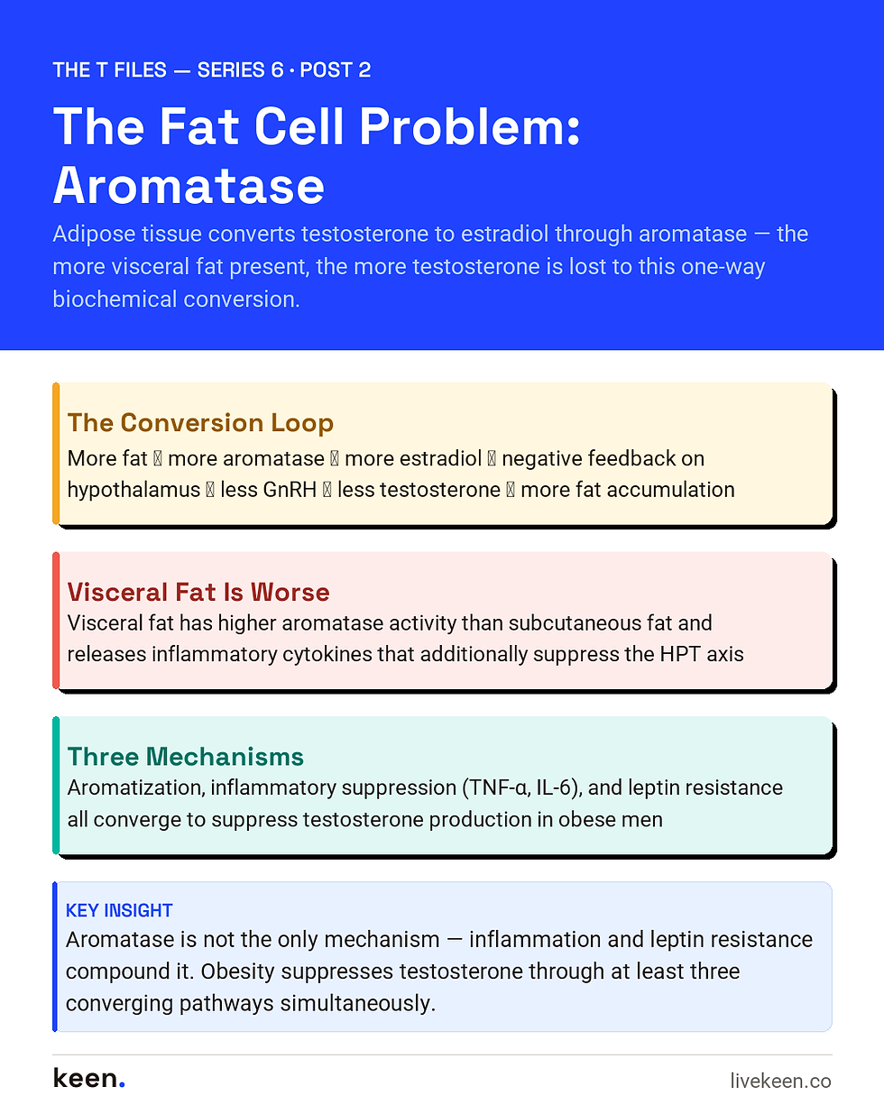
Your fat cells have a side hustle, and it is not the kind you want them to have.
Adipose tissue — body fat — is not simply inert storage. It is metabolically active. It secretes hormones, releases inflammatory cytokines, and expresses enzymes that alter the chemistry of the hormones circulating through your body. One of those enzymes is aromatase (formally, cytochrome P450 19A1, though nobody calls it that in conversation). And what aromatase does is convert androgens — primarily testosterone — into estrogens.
This is a normal biological process. Men produce small amounts of estradiol this way. A certain amount of estrogen is necessary for male bone health, cardiovascular function, and even sexual function. The problem is a dose-response relationship: the more adipose tissue you carry, the more aromatase activity you have, the more testosterone gets converted to estrogen, and the more your body’s control system interprets this as a signal to slow testosterone production down further.
The result is a biochemical feedback loop that gets worse the more body fat accumulates.
Aromatase: The Conversion Engine
The aromatase enzyme is expressed in multiple tissues, including the brain, bone, and gonads. But its expression in adipose tissue is what makes obesity a testosterone-suppressing condition. Adipose aromatase activity scales roughly with adipose tissue mass — more fat, more enzyme, more conversion.
The reaction itself is irreversible: aromatase catalyzes the conversion of testosterone (an androgen with 19 carbons) into estradiol (an estrogen with 18 carbons) by removing one carbon and aromatizing the A-ring of the steroid molecule. Once testosterone has been aromatized into estradiol, it cannot be converted back.
In men with high body fat, particularly those carrying significant visceral fat (the fat stored around internal organs and in the abdominal cavity), aromatase activity can become high enough to meaningfully reduce circulating testosterone while simultaneously elevating circulating estradiol.
This matters at the level of the hypothalamic-pituitary-testicular (HPT) axis because estradiol is one of the primary negative feedback signals in male testosterone regulation. When estradiol rises, the hypothalamus reduces its secretion of gonadotropin-releasing hormone (GnRH). GnRH is the upstream signal that drives the pituitary to release LH (luteinizing hormone) and FSH (follicle-stimulating hormone). LH, in turn, is the primary driver of testosterone synthesis in the Leydig cells of the testes.
So: more fat → more aromatase → more testosterone converted to estradiol → elevated estradiol suppresses GnRH → less LH released → less testosterone produced by the testes → lower testosterone levels → more fat accumulation (as testosterone normally promotes lean mass and opposes fat deposition) → more aromatase → repeat.
The Inflammatory Contribution
Visceral adipose tissue, in particular, has a troubling property beyond aromatase: when it is metabolically stressed — in the context of insulin resistance, which is common in obesity — it releases elevated levels of pro-inflammatory cytokines. These include tumor necrosis factor-alpha (TNF-α), interleukin-6 (IL-6), and others.
These cytokines suppress the HPT axis at multiple levels. They reduce GnRH pulsatility at the hypothalamus. They impair the pituitary’s gonadotropin response. And they may directly suppress Leydig cell function in the testes.
This means the obesity-aromatase hypothesis is not the only mechanism in play — it is one of several converging suppressive signals that happen to all be generated by the same metabolic state. Insulin resistance produces more inflammatory cytokines. More inflammatory cytokines suppress more of the HPT axis. And elevated insulin itself suppresses SHBG production by the liver, which changes the balance of free and bound testosterone in ways discussed further in the next post.
Grossmann and colleagues, in their 2014 review in Asian Journal of Andrology (PMID 24407187), noted that while the aromatase hypothesis has biological plausibility and clinical support — aromatase inhibitor use in obese men with hypogonadism does increase testosterone levels — the evidence from the EMAS suggested that even in non-diabetic obese men, estradiol levels were actually low, not high, and correlated positively with testosterone. This complicates the simple “aromatase converts T to E2, and high E2 suppresses the axis” narrative. Local tissue estradiol levels may not be fully reflected in circulatory measurements, and the inflammatory and leptin-mediated pathways of HPT suppression may be more dominant contributors in many obese men than the aromatase conversion pathway alone.
The honest picture is that obesity suppresses the HPT axis through multiple overlapping mechanisms, of which aromatase-mediated conversion is important but not the sole driver.
Leptin: The Appetite Hormone That Overstays Its Welcome
Leptin deserves a mention here, because it is another adipose-derived signal that connects obesity to HPT suppression. Leptin is a hormone secreted by fat cells in proportion to fat mass. It serves, in physiologically normal conditions, as a satiety signal to the hypothalamus — essentially a message saying “we have enough energy stored, you can reduce appetite.”
In severely obese individuals, however, the hypothalamus becomes resistant to leptin signaling, a phenomenon analogous to insulin resistance. The fat cells are still secreting leptin — often in very high amounts — but the brain has lost sensitivity to the signal. This leptin resistance also correlates with HPT axis suppression; the mechanisms are complex and involve crosstalk between leptin signaling pathways and GnRH pulsatility. Studies have shown that leptin receptors are expressed in Leydig cells, and supraphysiologic leptin concentrations can directly inhibit testosterone synthesis.
So the obese man’s adipose tissue is simultaneously: running an aromatase factory that converts testosterone to estrogen, releasing inflammatory cytokines that suppress his GnRH and LH output, and dumping leptin into his circulation at levels that have both central and peripheral testosterone-suppressing effects.
This is not a mild situation requiring minor adjustments. It is a systemic metabolic environment that is inhospitable to testosterone production at every level of the axis.
What This Looks Like Clinically
The clinical presentation of obesity-associated functional hypogonadism is not always distinct from other causes of low testosterone. Men present with fatigue, reduced libido, difficulty maintaining erections, mood changes, and reduced motivation. Lab work shows low or low-normal testosterone, often with low or inappropriately normal LH — the pattern of secondary hypogonadism.
What the clinical picture often also includes: central obesity, elevated fasting glucose or frank insulin resistance, elevated blood pressure, and elevated triglycerides — the metabolic syndrome cluster. These are not coincidental co-presentations. They are mechanistically connected to the same hormonal disruption.
The clinical implication — which will be explored further across this series — is that in a man whose hypogonadism is predominantly driven by obesity, the most targeted intervention for the underlying cause is weight reduction. But the biology also makes weight reduction particularly difficult in hypogonadal men. Which is precisely the trap this series is about.
Next up: Post 3 — “SHBG and the Binding Problem: Why Insulin Resistance Changes What Your Free Testosterone Number Actually Means.”


